|
|
|
Keynote speakersFIMH 2019 will feature four keynote speakers:
Title: Building the Faith: Validating Computational Models for Clinical Use
Abstract: Computational modeling has been an integral part in the advancement of our understanding of heart function along with experimental and theoretical approaches. Physiological mechanistic cardiac models predict macroscopic phenomena such as electrical impulse propagation and contraction throughout the entire heart as well as flow and pressure dynamics occurring in the ventricular chambers, aorta, and coronary arteries. Such models have been used to study a variety of clinical scenarios including aortic aneurysms, coronary stenosis, cardiac valvular disease, left ventricular assist devices, cardiac resynchronization therapy, ablation therapy, and risk stratification. After decades of research, these models are beginning to be incorporated into clinical practice directly via marketed devices. However, if models are to be used in safety-critical clinical decision making, the reliability of their predictions needs to be rigorously and comprehensively investigated. In engineering and the physical sciences, the field of ‘verification, validation and uncertainty quantification’ (VVUQ) (also known as ‘verification and validation’ (V&V)) has been developed for rigorously evaluating the credibility of computational model predictions. In this talk I will discuss the importance of the VVUQ framework in model development and evaluation and FDA’s efforts to enable these potentially transformative computational models to enter clinical practice. First, I will present unique challenges to VVUQ for modeling of physiological phenomena. Then I will present various aspects of VVUQ using our current research to demonstrate the ideal and practical aspects in the field of cardiac electrophysiology which include calibration, parameter sensitivity, model complexity, and context of use of a model. Overall, the feasibility of performing VVUQ for cardiac cell models, and the assessment of model robustness, will be demonstrated. Finally, I will stress the need of rigorous evaluation whose aim is to increase model robustness for computational clinical tools with the goal of enabling reliable use in safety-critical decision-making.
Short biosketch: Richard A. Gray, PhD is a Senior Research Biomedical Engineer at the FDA where he leads the Computer Modeling Lab and is developing methods for verification, validation, and uncertainty quantification of complex computer physiological models. He also evaluates new medical device technology and performs regulatory science applied research to develop new methods to rigorously evaluate complex device related issues related to cardiac pacemakers, defibrillators, advanced algorithms, and mapping systems. Previous to FDA he was faculty and Director of the Biomedical Engineering Graduate Program at the University of Alabama at Birmingham. His full curriculum vitae includes over 150 peer reviewed publications, 35 invited presentations, Principal Investigator on 7 Grants (including NIH, and NSF), as well as numerous awards, positions and significant intra- and extramural servicee
Title: Electromechanical wave imaging for nonivasive and direct mapping of arrhythmia in 3D.
Abstract: Arrhythmias refer to the disruption of the natural heart rhythm. This irregular heart rhythm causes the heart to suddenly stop pumping blood. Arrhythmias increase the risk of heart attack, cardiac arrest and stroke. Reliable mapping of the arrhythmic chamber stands to significantly improve these currently low treatment success rates by localizing arrhythmic foci before the procedure starts and following progression throughout. To this end, our group has pioneered Electromechanical Wave Imaging (EWI) that characterizes the electromechanical function throughout all four cardiac chambers. The heart is an electrically driven mechanical pump that adapts its mechanical and electrical properties to compensate for loss of normal mechanical and electrical function as a result of disease. During contraction, the electrical activation, or depolarization, wave propagates throughout all four chambers causing mechanical deformation in the form of the electromechanical wave. This deformation is extremely rapid and completes within 15-20 ms following depolarization. Therefore, fast acquisition and precise estimation is extremely important in order to properly map and identify the transient and minute mechanical events that occur during depolarization. Our group has demonstrated that EWI yields 1) high precision electromechanical activation maps that include transmural propagation, 2) imaging of transient cardiac events (electromechanical strains within ~0.2-1 ms). Our studies have shown the EWI capable of capability atrial fibrillation and atrial flutter, transmural atrial pacing, RF ablation lesions while more recently it has been shown more robust that 12-lead EKG in characterizing focal arrhythmias such as the Wolf-Parkinson-White (WPW) and Pre-ventricular Contraction (PVC) as well as macro-reentrant arrhythmias in patients. The most relevant recent findings of the aforementioned studies will be presented. Short biosketch: Prof. Konofagou received her B.Sc. (Licence) in chemical physics from the Paris VI University (Université de Pierre et Marie Curie; Paris, France) and her M.Sc. in biomedical engineering from Imperial College (London, UK) in 1992 and 1993, respectively. In 1999, Prof. Konofagou received her Ph.D. in biomedical engineering from the University of Houston (Houston, TX, USA) for her work in elastography at the University of Texas Medical School. She then carried out postdoctoral work in elasticity-based monitoring of focused ultrasound therapy at Brigham and Women's Hospital (Boston, MA, USA), an affiliate of the Harvard Medical School. Prof. Konofagou is currently a Professor of Biomedical Engineering and Radiology and is also the Director of the Ultrasound and Elasticity Imaging Laboratory at Columbia University. She is a member of the IEEE Ultrasonics, Ferroelectrics and Frequency Control group; the Acoustical Society of America; and the American Institute of Ultrasound in Medicine. Prof. Konofagou's main interests are in the development of novel elasticity imaging techniques and applications, such as breast elastography, ligament elastography, electromechanical wave imaging (EWI), myocardial elastography, harmonic motion imaging, pulse wave imaging (PWI), and focused ultrasound therapy, in particular research on the blood-brain barrier opening. Prof. Konofagou maintains several close clinical collaborations in the Columbia Presbyterian Medical Center.
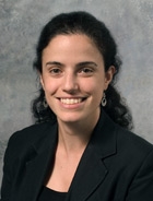
Title: Patient-specific hemodynamics simulations for interventional planning of congenital and acquired cardiac diseases Abstract: Hemodynamics modeling has become mature enough to simulate local fluid dynamics changes due to a surgery or device implantation. However taking into account their interactions with the rest of the circulation, or even the downstream vascular bed not accessible by imaging remains challenging. We will present several computational methods that have been developed to tackle this issue for patient-specific image-based modeling. We will demonstrate through several examples of acquired and congenital heart disease how such simulations can be performed by including morphological and functional data. We will also illustrate what they can bring in this context: for example, by testing separately or all together, the effects of disease worsening and intervention, or by being the basis for a risk predictor. Short biosketch: Irene Vignon-Clementel, PhD is Directeur de recherche (Prof. equiv.) at Inria, Mathematics & Informatics. She holds a ‘habilitation’ degree in Applied Mathematics (UPMC), a PhD in mechanical engineering (Stanford University), an MS in Applied Mathematics (DAMTP, Cambridge U.) and a diplôme d’ingénieur from Ecole Centrale Paris (France). Her research focuses on modeling and numerical simulations of physiological flows to better understand certain pathophysiologies and their treatment (surgical planning, medical device design), especially around blood circulation and breathing. This requires developing models of different complexities, coupling them, that their numerical implementation is robust, and that their parameters are based on medical or experimental data specific to a subject. Applications include congenital cardiac diseases, liver (surgery, remodeling), emphysema and asthma, and more recently the interpretation of non-invasive dynamic imaging (MRI, fluorescence, etc.). She has written over 70 publications and several book chapters, is member of several conference committees and of the IJNMBE editorial board. She received the top recipient award of the western states American Heart Association fellowship (2004-2006), the student award at the World Congress of Computational Mechanics by the USACM and the USACM Executive Committee (2006), and Inria excellence award (2012 and 2016). Dr. Vignon-Clementel has been working with companies and clinicians as a PI in a number of national and international grants (such as a Leducq transatlantic network of excellence), and actively promoting the applied mathematics/bioengineering and medicine interface through co-supervision of MD-PhDs, joint research projects, conference organization and interface articles with clinicians.
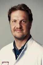
Title: Image-based Modeling for the Clinical Management of Patients with Cardiac Arrhythmias: Are We There Yet? Abstract: Over the past 2 decades, paralleling several major developments of cardiac interventions in structural heart disease, cardiac imaging has evolved from a diagnostic use to a decision-making tool employed to tailor patient management and assist interventions. In the context of cardiac arrhythmias, the use of imaging to personalize computational models of cardiac electrophysiology has been proposed to extract physiological markers with potential clinical applications for either patient selection/risk stratification or therapy optimization/ablation targeting. In this lecture, will present a clinician perspective on these new developments. I will summarize the current methods and indications for cardiac imaging in the context of cardiac arrhythmias, the remaining clinical issues that image-based computational models may address, and the challenges of implementing such approaches in clinical practice. Short biosketch: Hubert Cochet, MD, PhD, is Professor of Radiology and Medicine at University of Bordeaux, head of the cardiac imaging clinic at Bordeaux University Hospital, and senior researcher at IHU Liryc, a multidisciplinary research institute focusing on cardiac electrical disorders. His research consists of combining imaging and electrophysiological data to better understand the mechanisms involved in cardiac electrical diseases. It aims at developing image-based strategies for improved diagnosis, risk stratification and therapy guidance in these extremely prevalent diseases. Along with Pierre Jais (Cardiologist) and Maxime Sermesant (Computer Scientist), Hubert Cochet has developed software for patient-specific image-based modeling that is now used on a daily basis to guide catheter ablation procedures in over 30 centers worldwide. Pr Cochet has authored more than 100 publications in major Cardiology and Radiology journals, and is involved in multiple National and European-funded research projects, including as PI of a recently-awarded ERC grant to conduct research at the interface between cardiac imaging and electrophysiology. |
| Online user: 1 | RSS Feed |

|


